г. Санкт-Петербург, ул. Ярослава Гашека, 21. Телефон: +7 (812) 324-66-66
Glaucoma is one of the leading causes of blindness in the world. This disease is characterized by changes in the optic nerve which leads to restriction of visual field and even blindness. These changes can develop so slowly, that You will not notice them until there are some irreversible damages of the optic nerve.
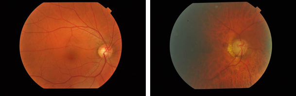
On the left You can see an example of a normal fundus of the eye: optic nerve disk is of pale pink color with physiological excavation, on the right You can see fundus of the eye with advanced glaucoma: disk of the optic nerve is grey with deep excavation
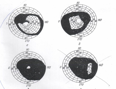
Changes to the visual field in glaucoma case starts with nasal side, while the process is progressing visual field is concentrically reducing, at terminal stages only tunnel vision is kept or periphery visual field island
Normal vision
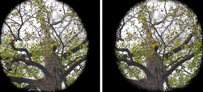
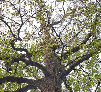
Left picture shows how the left eye with advanced glaucoma sees, the right picture shows how the right eye with advanced glaucoma sees
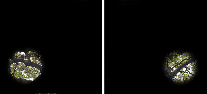
On the left is an example of almost terminal glaucoma stage vision for the left eye, right picture shows the same for the right eye.
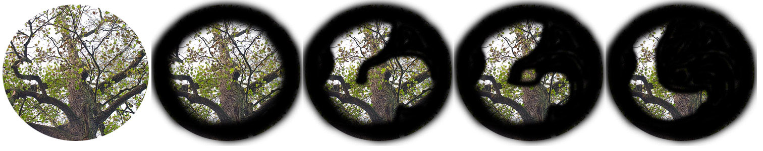
Development of visual field reduction in case of glaucoma
One of the glaucoma symptoms is high intraocular pressure.
Early diagnostics and treatment can make less or prevent the optic nerve damages. It is important to visit Your doctor systematically and get Your intraocular pressure checked.
There are two main types of glaucoma – open angle and angle-closure glaucoma.
Open angle glaucoma happens more often (90-95%) and is more cunning because it may progress almost without any symptoms until the vision is completely lost. In case of this glaucoma type visual field is narrowing gradually, and if the second eye has a good visual acuity, You will possibly not even notice it until You are blind.
Angle-closure glaucoma occurs more seldom and is accompanied by halo periodically appearing around the light sources, foggy vision, in some cases a sharp ocular pain, eyes redness, sudden vision loss, nausea and vomiting.
Do not hesitate and wait until You have obvious visual problems, any inexplicable visual discomfort requires visiting Your ophthalmologist!
Regular examinations by Your ophthalmologist are the key to finding glaucoma at early stage, which helps to treat the disease and prevent its further development. It is recommended to have a complex eye examination every 3-5 years for all people above 40. You should be more aware if one of your relatives had glaucoma or blindness with no particular reason. Apart from that, mind that a strong headache or ocular pain, nausea, foggy vision or halo around sources of light might be also symptoms of acute glaucoma attack. If you have any of these symptoms or all of them, consult ophthalmologist immediately.
Mostly glaucoma may have no any noticeable symptoms until the disease doesn’t cause irreversible damages. If not treated, it can lead to a progressive vision loss. At terminal stages glaucoma damages are practically irreversible even in case of successful intraocular pressure decrease.
Regular complex examinations will help to find glaucoma at early stages, when irreversible changes haven’t happened yet. Usually, it is recommended to have a complex eye examination every 3-5 years for all people above 40 and every year after 60. More often screening may be needed if You have any of the risk factors for glaucoma development.
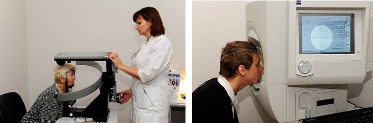
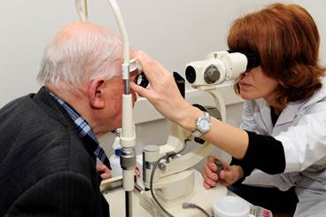
For glaucoma diagnostics and efficient treatment You need to make the following tests:

Mainly glaucoma is treated by decreasing the intraocular pressure in order to decompress the optical nerve. Glaucoma is a chronic disease, that’s why full recovery is impossible and the visual acuity that is lost usually is not restored. But diagnostics and treatment performed in the right time, as well as regular checkups may prevent the vision loss, and if it did happen already then it can slow down the further process or even prevent from going blind.
Glaucoma treatment often starts with prescription of eye drops. There are many drugs now that can make intraocular pressure come down and cut the risks of glaucoma progressing. These eye drops must be used without any pause and their efficiency should be checked up by a doctor.
If eye drops do not decrease the intraocular pressure to the necessary level, they can be replaced with pills (inhibitors of carbonic anhydrase), but their long-term usage may cause side effects including hypokalemia, paresthesia, muscle weakness, seizure, skin hyperemia, itching, buzzing in the ears, nausea, vomiting, metabolic acidosis, vesiculation, urinary frequency, urolithiasis exacerbation.
At present a whole system of different laser glaucoma surgeries for different types of glaucoma is developed and put into practice. It allows to choose an adequate method in each case.
Main methods of laser surgery for glaucoma:
It is necessary to pay attention that laser and other surgery performance doesn’t guarantee full recovery from glaucoma, which is why the patient should continue to visit his ophthalmologist as he did before. This is necessary not to miss a rising intraocular pressure that rises, due to the scar process after the outflow paths which were formed as a result of laser or surgical intervention were closed.
Surgical treatment is the most efficient way of decreasing the intraocular pressure.
The purpose of surgical treatment is not to improve the visual acuity, but to keep the vision you have by decreasing the intraocular pressure, that is why it is very important to perform glaucoma surgery at early stages while irreversible damages to the optic nerve didn’t happen.
When considering the time period for surgical treatment, it should be kept in mind, that modern strategy of glaucoma patients management is mainly focused on early stages surgery providing better prognosis and safety. Recent studies have shown that the course of operated glaucoma is more favorable than the course of non-operated.
При выполнении операции формируются дополнительные пути оттока внутриглазной жидкости, что способствует нормализации внутриглазного давления.
It should be noted that after performed surgery patient requires a constant supervision by ophthalmologist, since in most cases glaucoma develops on the second eye, and in the long term postoperative period, as a result of scarring in the formed outflow pathways, intraocular pressure may rise on the operated eye, which can require repeated surgical interventions.
In conclusion, it is necessary to remind, that regular complex examinations may help to detect glaucoma at early stages, when irreversible changes didn’t come yet. Usually it is recommended to have a complex eye examination every 3-5 years for all people above 40 and every year after 60. More often screening is needed in case you have any of the risk factors.
Примите, пожалуйста, участие в опросе о качестве нашего сайта